

iWorks FX
Microscope Software for Particle Analysis & Automatic Counting
iWorks FX is the outstanding microscope software for automatic objects or cells counting, particle analysis, fluorescence Image multiple color merge and metallurgical area fraction. It is a good choice for high-end biological specialist and users of metallographic analysis.
Note: All functions of iWorks LT are included in iWorks FX.
Manual Counting
Manual counting tool, by mouse clicking.

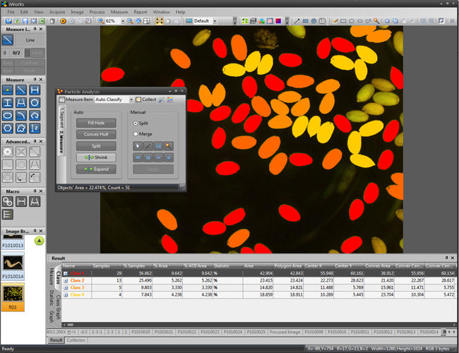
Automatic Counting / Particle Analysis
When measurements are made, the program highlights the measuring outlines and assigns reference numbers to the objects in your image. iWorks introduce to advanced particle analysis gets an object by simple click of your mouse then measure and analysis all of object by automatically.
AOI (Area of Interest) Manager
Define your prefered area with several tools.


Enhanced Image Processing Tools
-
Image enhancement: Invert, Brighteness, Contrast, Gamma
-
Arithmetic/ Logic operation
-
Color split/ merge
Merge: Generate one or several R/G/B grayscale images to a new color image.

Split: Used for extracting one or several R/G/B channels from a color image.

-
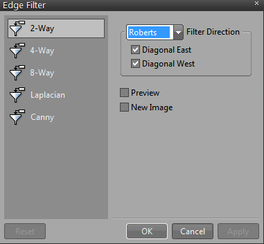
Image Filters (Spatial Filter, Edge Filter)

-
Histogram

Segmentation

Fluorescence Manager and Pre-defined Dye List
Multiple single channel images (captured with different optical filters or under different camera settings) can be merged together simply by using Fluorescence Manager of iWorks. The combined images can be stored to a file while maintaining their original bit depths or optionally can be converted into a RGB image.


